
Salivary Gland Disorders

Each person has six major and many minor salivary glands which produce saliva to help lubricate the mouth and the throat. The major salivary glands are the parotid glands (located in your cheek), the submandibular glands (located beneath your jaw), and the sublingual glands (located beneath your tongue). Each person has two of these glands, one on each side of the body.
When a salivary gland becomes inflamed, it enlarges and becomes painful. This can happen because of dehydration, bacterial infection, or even salivary stones. Often times, hydration, antibiotics, and steroids are all that is needed to resolve salivary gland inflammation. The salivary glands produce stones similar to your kidneys, and when these salivary stones form in the ducts which drain the glands, the treatment of choice is to have the stone removed.
Removing the stone can sometimes be performed in the office under local anesthesia, or through salivary endoscopy where small telescopes are used to navigate the ducts and remove the stones. Occasionally, the entire gland must be removed through surgery.
Salivary glands can also have abnormal growth of cells called neoplasms, which can be either benign or malignant. These growths occur most frequently in the parotid gland, and 75-80% of these growths are benign. When a salivary neoplasm occurs in other locations, the risk of malignancy is higher. With a few exceptions, the recommended management of salivary gland neoplasms is surgical excision. Minimally invasive parotidectomy techniques allow removal of parotid tumors with no visible scarring on the face or neck.
Salivary Stones
Salivary stones form in the ducts of the salivary glands located in the cheeks (parotid glands) or beneath the jaw (submandibular glands). By themselves, they are not painful and at times are asymptomatic. Most stones are very small, on the order of a few millimeters in size. Stones are sometimes diagnosed clinically, and on CT scan, and they show up as white spots that look like bone.
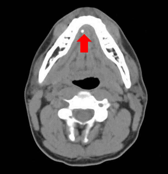
This image shows a 3 mm stone in the right submandibular duct.
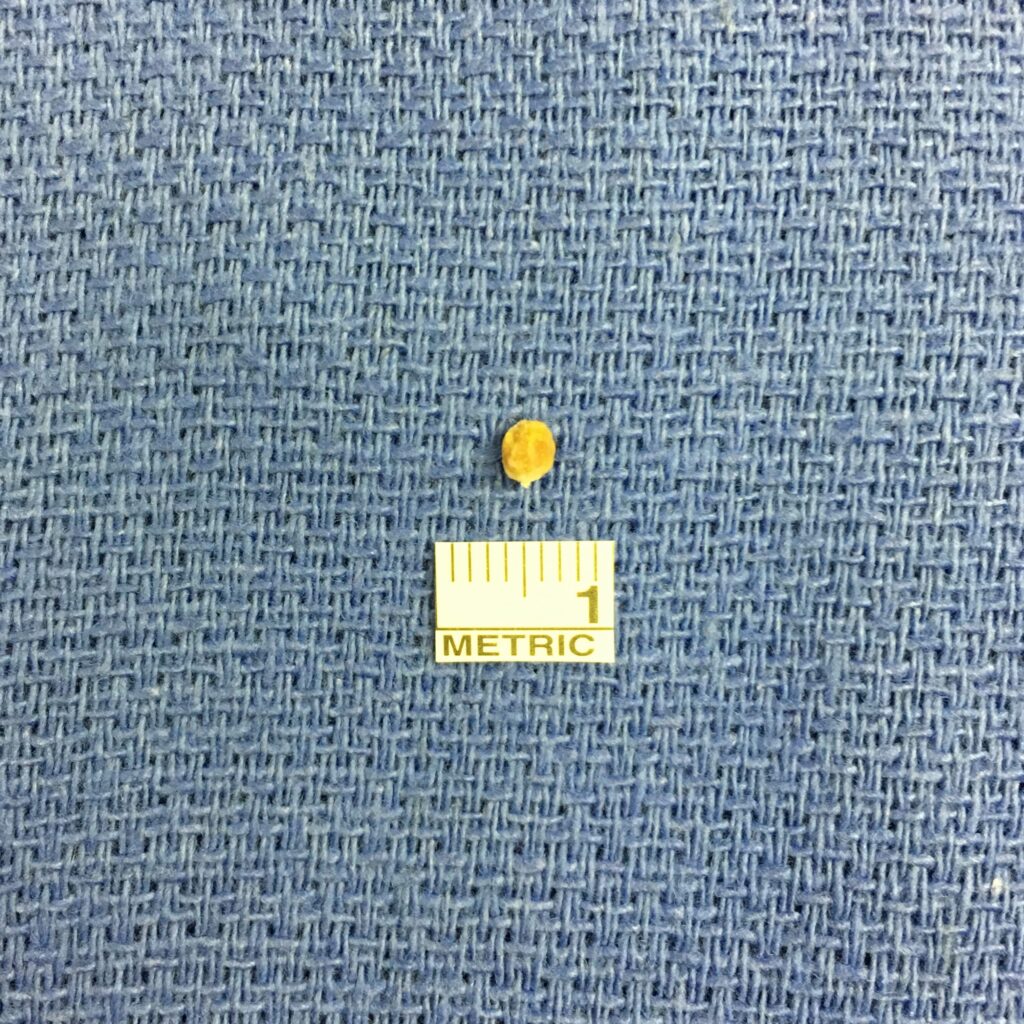
Here is the stone after it was removed through salivary endoscopy, beneath a ruler which is 1 cm in length.
When salivary stones obstruct the flow of saliva, the pressure backs up into the salivary gland and causes inflammation of the gland, pain, and even infections. Many times, symptoms can be improved with use of warm compresses, gentle massage, and use of sour candies which encourages the salivary glands to produce more saliva. Symptoms may improve over time, but the definitive management of salivary stones is removal. Removing a stone can sometimes be performed in the office under local anesthesia, or through salivary endoscopy where small telescopes are used to navigate the ducts and remove the stones. Occasionally, the entire gland must be removed through surgery.
Salivary Endoscopy
Salivary endoscopy, or sialendoscopy, is a minimally invasive technique for treating disorders of the salivary ducts. The most common indication for salivary endoscopy is for removal of salivary stones. Historically, the only treatment for salivary stones was to surgically remove the affected salivary gland. However, the recent development of salivary endoscopes and small tools which fit through the scope has allowed minimally invasive removal of stones without removing the gland.
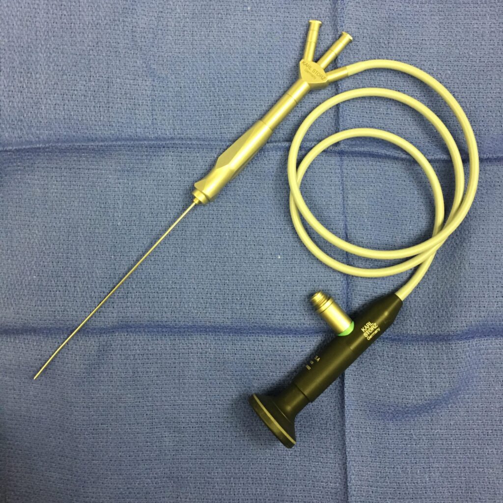
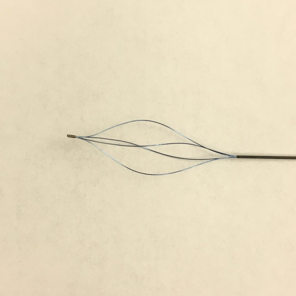
This is a salivary endoscope which is introduced into the salivary ducts to allow visualization and removal of salivary stones.
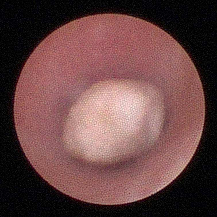
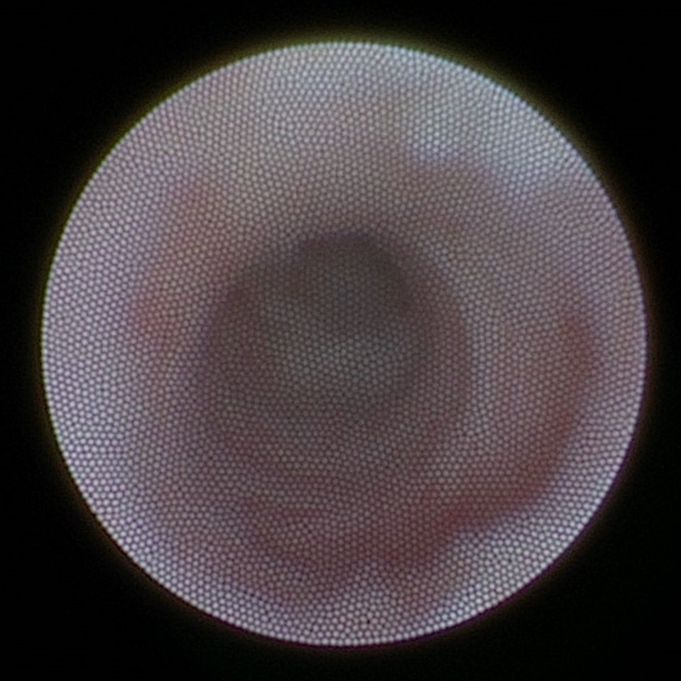
Here is the image of a salivary stone in one of the salivary ducts.
This is an image of the small retrieval basket which is inserted through the scope to remove salivary stones.
This is an image of a normal salivary duct after stone removal. It is widely patent after the stone has been removed. The diameter of this duct is only about 3 mm in size.
This procedure is performed on an outpatient basis, leaves no scars on the face or neck, and patients are back at work the next day without the need for pain medication. Few surgeons in Atlanta are currently performing this procedure.